Bladder exstrophy
The exstrophy-epispadias complex comprises a spectrum of congenital abnormalities that includes classic bladder exstrophy, epispadias, cloacal exstrophy, and several variants. Each of these anomalies is thought to result from the same embryologic defect. Separation of the primitive cloaca into the urogenital sinus and hindgut occurs during the first trimester at approximately the same time as maturation of the anterior abdominal wall. Failure of mesenchyme to migrate between the ectodermal and endodermal layers of the lower abdominal wall leads to instability of the cloacal membrane. Premature rupture of the membrane before its caudal translocation leads to this complex of infraumbilical anomalies. Rupture of the cloacal membrane after complete separation of the genitourinary and gastrointestinal tracts results in classic bladder exstrophy.
Defects
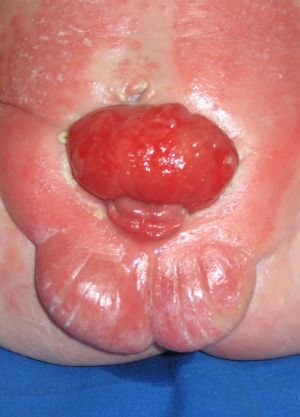
Classic bladder exstrophy. The bladder is open on the lower abdomen, with mucosa fully exposed through a triangular fascial defect. The abdominal wall appears long because of a low-set umbilicus on the upper edge of the bladder plate. The distance between the umbilicus and anus is foreshortened. Rectus muscles diverge distally, attaching to the widely separated pubic bones. Indirect inguinal hernias are frequent (>80% of males, >10% of females) due to wide inguinal rings and the lack of an oblique inguinal canal. The phallus is short and broad with upward curvature (dorsal chordee). The glans lies open and flat like a spade, and the dorsal component of the foreskin is absent. The urethral plate extends the length of the phallus without a roof. The bladder plate and urethral plate are in continuity, with the verumontanum and ejaculatory ducts visible within the prostatic urethral plate. The anus is anteriorly displaced with a normal sphincter mechanism.
Epispadias. The pubic symphysis is generally widened. The rectus muscles are divergent distally. (see Epispadias)
Cloacal exstrophy. Nearly all patients have an associated omphalocele. The bladder is open and separated into two halves, flanking the exposed interior of the cecum. Openings to the remainder of the hindgut and to one or two appendices are evident within the cecal plate. Terminal ileum may prolapse as a “trunk” of bowel onto the cecal plate. The penis is generally quite small and bifid, with a hemiglans located just caudal to each hemibladder. Infrequently, the phallus may be intact in the midline. In females, the clitoris is bifid and 2 vaginas are present. The anus is absent.
Exstrophy variants. The pubic symphysis is widely separated, and rectus muscles diverge distally. The umbilicus is low or elongated. A small superior bladder opening or a patch of isolated bladder mucosa may be present. The intact bladder may be externally covered by only a thin membrane. Isolated ectopic bowel segments have been reported. Patent urachus is a differential diagnosis for the superior vesical fissure variant of exstrophy-epispadias. However, patent urachus lacks the typical musculoskeletal abnormalities of exstrophy and is open at the umbilicus. Superior vesical fissure is infraumbilical. Genitalia generally are intact, though epispadias can occur.
Musculoskeletal defects
The pubic symphysis is widely separated. Divergent rectus muscles remain attached to the pubis. External rotation of the innominate bones results in a waddling gait in ambulatory patients but does not appear to result in orthopedic problems later in life.
Neurologic defects
In cloacal exstrophy, as many as 95% of patients have myelodysplasia, which may include myelomeningocele, lipomeningocele, meningocele, or other forms of occult dysraphism. These patients are at risk of neurologic deterioration, and they should be observed closely. Early neurosurgical consultation is appropriate if a radiographic abnormality of the spinal cord or canal is observed.
Treatment
Reconstruction of exstrophy-epispadias complex remains one of the greatest challenges facing the pediatric urologist. Many modifications in surgical procedures have improved the outcome for these patients, but the optimal approach remains uncertain. Longitudinal prospective assessment of both surgical approaches (staged procedure versus total reconstruction) is critical to optimize functional and cosmetic outcomes.
Goals of therapy include provision of urinary continence with preservation of renal function and reconstruction of functional and cosmetically acceptable genitalia. Creation of a neo-umbilicus is also important to many of these patients.
Surgical techniques include the following:
1. Staged functional closure for classic bladder exstrophy
Series of operations.
– Initial bladder closure is completed within 72 hours of birth. If delayed, pelvic osteotomies are required to facilitate successful closure of the abdominal wall and to allow the bladder to lie within a closed and supportive pelvic ring.
– Epispadias repair with urethroplasty at age 12-18 months allows enough increase in bladder outlet resistance to improve the bladder capacity.
– Bladder neck reconstruction at age 4 years (typically a modified Young-Dees-Leadbetter repair) allows continence and correction of vesicoureteral reflux. Multiple modifications have been proposed.
2. Complete primary repair for classic bladder exstrophy
– This is a relatively newer approach to exstrophy closure.
– Primary bladder closure, urethroplasty, and genital reconstruction are performed in a single stage in newborns. This procedure involves complete penile disassembly in males and mobilization of the urogenital complex in females. Hypospadias is a common outcome in males and requires subsequent reconstruction.
– The goal is early bladder cycling. A subset of patients has achieved continence without bladder neck reconstruction
I am text block. Click edit button to change this text. Lorem ipsum dolor sit amet, consectetur adipiscing elit. Ut elit tellus, luctus nec ullamcorper mattis, pulvinar dapibus leo.
CASE 1
Extrophy epispadias complex repair
Extrophy epispadias complex in male newborn

One stage repair: Bladder reconstruction and penile reconstruction
CASE 2
Extrophy epispadias complex repair

Newborn with extrophy epispadias complex

Newborn with extrophy epispadias complex

Penile disassembly / bladder dissection

Penile disassembly / bladder dissection

Bladder closure

Bladder closure

Final appearance

Final appearance