Penile fracture
Penile fracture is a traumatic rupture of tunica albuginea and the tumescent corpora cavernosa due to the nonphysiological bending of the penile shaft, presenting with or without rupture of corpus spongiosum and urethra. It is relatively uncommon injury, occurring approximately 1 in 175000 hospital care emergencies, not including notable number of patients not seeking for medical care due to embarrassment or fear.
Etiology
As a result of the trauma of an erect penis, the most common mechanism of penile fracture is during sexual intercourse – the penis slips out of the vagina and strikes the perineum or pubic symphysis. Other factors include: masturbation, penile manipulation to achieve detumescence, direct blow to the erect penis, as well as many other types of trauma of penis in the erect state.
Pathophysiology
The tunica albuginea is the fibrous covering of the penile corpora cavernosa and is directly involved in maintaining an erection. During erection it thins from 2 mm to 0.25 – 0.5 mm, stiffens and becomes less elastic. Sudden direct trauma or abnormal bending in erect state causes tearing of the tunica albuginea, usually in transversal direction, rarely oblique or irregular. Corpora cavernosa becomes exposed to trauma, leading to fracture of one corpus, rarely both corpora cavernosa. Consequent urethral injury or rupture of corpus spongiosum must precisely be assessed and repaired.
Urethral injury
Urethral injury is associated in 10-30% of cases of penile fracture. It can manifest by the appearance of blood at the urethral meatus or in urine (hematuria), as well as dysuria and urinary retention. Injury of the urethra can also remain unrecognized as being asymptomatic.
Rupture of the deep dorsal vein
Rupture of the deep dorsal vein has similar manifestations as penile fracture, and surgical exploration is necessary to differentiate. Swelling and ecchymosis are present, without typical crack or popping sound, and without rapid development of detumescence.
Clinical presentation
Characteristic snapping or popping sound occurs, followed by sharp penile pain and rapid detumescence. Penile deformity, swelling and discoloration (ecchymosis) due to leakage of the blood from corpus cavernosum, are visible. Typically patients report sharp pain and cracking sound, with consecutive loss of erection, deformity and discoloration.
Medical history and physical findings are enough to confirm the diagnosis in most cases. Imaging studies in diagnosis of penile fracture are required only when injury is not evident on physical examination. Retrograde urethrography, penile cavernosography or magnetic resonance imaging can be used in those cases.
Treatment
Conservative treatment was considered as treatment of choice in the past. It includes cold compresses, pressure dressing, and administration of anti-inflammatory drugs, fibrinolytics, anti-androgens and sedatives. It results in complications in 29 – 53% of penile fracture cases. The most common complications of conservative treatment are missed urethral injury, penile abscess, permanent curvature, presence of the nodule formation at the site of the fracture (fibrotic plaque), fistulas (arteriovenous, corporourethral), as well as painful erection and erectile dysfunction.
Immediate surgical repair in case of penile fracture is required. The aim of surgical correction is penile restoration in preinjury state, prevention of penile deformities and erectile dysfunction, maintenance of penile length and retrieving of normal voiding. Surgical treatment is based on several principles due to complete assessment of the injury and penile restoration in previous state:
– optimize the surgical exposure:
* direct incision over the defect
* circumscribing-degloving incision
* inguinal-scrotal incision
– evacuate the hematoma
– identify the site of injury
– correct the defect of tunica albuginea
– repair the urethral injury
CASE 1
Small penile fracture involving the left corpus cavernosum

Penile deformity, swelling and discoloration due to fracture

Penile deformity, swelling and discoloration due to fracture

Intraoperative appearance

Repair of the left corpus cavernosum
CASE 2
Severe penile fracture

Appearance of the penis after severe fracture
Intraoperative appearance of the severe fracture
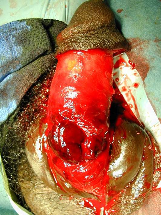
CASE 3
Severe penile fracture with “eggplant” appearance

Blood at the meatus – urethral injury

Blood at the meatus – urethral injury

Marked rupture of the left corpus cavernosum associated with urethral rupture

Repair of the corpora cavernosa. Closure of the urethral defect.

Postoperative dressing
CASE 4
Penile fracture repair two months after injury

Appearance of the penis two months after fracture

Echosonographic appearance – hematoma is close to the right corpus cavernosum

Diverticula formation at the site of the fracture

Diverticula formation at the site of the fracture

Removal of the diverticula

Removal of the diverticula. Reconstruction of the tunica albuginea.
CASE 5
Urethral stricture after previously treated penile fracture

Narrowing of the penis at the site of the fracture

Buccal mucosa graft – dorsal “roofing”

Buccal mucosa graft – dorsal “roofing”

Buccal mucosa graft – dorsal “roofing”

Repair of the tunica albuginea by penile skin graft
1. Djordjevic ML. Surgery: Treating urotrauma – new guidelines to aid decisions. Nat Rev Urol. 2014;11(9):495-6.
2. Gómez RG, Storme O, Catalán G, Marchetti P, Djordjevic M. Traumatic testicular dislocation. Int Urol Nephrol. 2014 May 29. [Epub ahead of print] PMID:24869967
3. Perovic SV, Djinovic RP, Bumbasirevic MZ, Santucci RA, Djordjevic ML, Kourbatov D. Severe penile injuries: a problem of severity and reconstruction. BJU Int. 2009, 104(5):676-87.
4. Djordjevic M, Santucci R, Bumbasirevic M, Perovic S. Outcomes and special techniques for treatment of severe penile injuries. Urology, 2007; 70(suppl. 3a):167(abs.MP22.06).