Peyronie’s disease
Peyronie’s disease (also known as plastic induration of the penis) is characterized by the formation of hardened tissue (fibrosis) in the penis that causes pain and bending of the penis, affecting up to 10% of men. Although the popular conception of Peyronie’s disease is that it always involves curvature of the penis, the scar tissue sometimes causes divots or indentations rather than curvature. Progression of the disease can lead to shrinking and shortening of the penis and erectile dysfunction. The condition may also make sexual intercourse painful and/or difficult.
Etiology
The cause of Peyronie’s disease is unknown, but it is generally accepted that minor repetitive trauma can be an etiological factor. Abnormal healing can result in development of hard, thickened scar tissue (plaque) in the tunica albuginea. With repetitive trauma, the plaque may develop tough fibrous tissue (fibrosis) or calcium deposits (calcification) and result in deformity. Recent studies show that tunica albuginea is the main factor of erection, implying that multifocal deformities of tunica lead to erectile dysfunction.
Clinical manifestation
Progression of the disease initially brings pain and various penile sensations (paresthesia), then formation of plaques, finally resulting in deformity (curvature), penile retraction and erectile dysfunction. In most cases there are four main symptoms: painful erection, penile deformities or penile shortening in erection, presence of plaque or indurations on penile corpus, and erectile dysfunction.
Physical examination can confirm presence of hard plaque inside the penis. It may be necessary to inject medication (prostaglandin E1) into the penis to induce an erection for proper evaluation.
Treatment
Conservative treatment
Conservative treatment, such as oral medications, local treatment and intralesional injections, is usually poorly effective, particularly if severe deformity is present.
Surgical treatment
Surgical treatment is required in most cases. Preoperative evaluation of penile vascularisation and erectile (dys)function is necessary. There are three options for surgical treatment:
1. “tunical lengthening procedures”,
2. procedures including implantation of penile prosthesis,
3. “tunical shortening procedures”.
First option implies incising the concave side of corpora cavernosa and grafting the defect, and the second one – excising the plaque and implanting the penile prosthesis with or without grafting. Surgical approach depends on presence of erectile dysfunction and severity of disease. We avoid using plication techniques since they can provoke further shortening of the penis. In patients with preserved erection our preference is grafting procedure that guarantees satisfactory result in straightening and lengthening of the penis. Using precise geometrical principles in creating and fashioning the graft with appropriate size leads to precise correction with penile lengthening. Patients with erectile dysfunction that is not responding to conservative treatment are candidates for implantation of penile prosthesis. Postoperative treatment entails physical therapy by vacuum device which is of crucial importance. If significant erectile dysfunction is present, simultaneous implantation of penile prostheses is performed to obtain suitable penile rigidity.
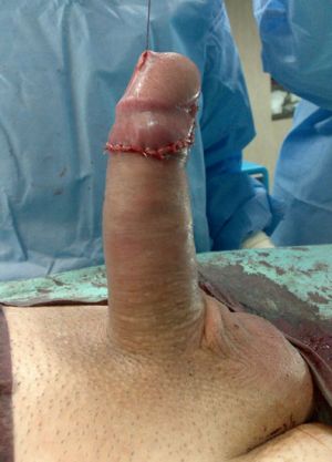
CASE 1
Peyronie’s disease – grafting technique

Severe deformities confirmed intraoperatively by artificial erection.

Severe dorsolateral curvature.

Correction of all deformities is done using bovine pericardium graft.

Penis is completely straightened using grafting technique.
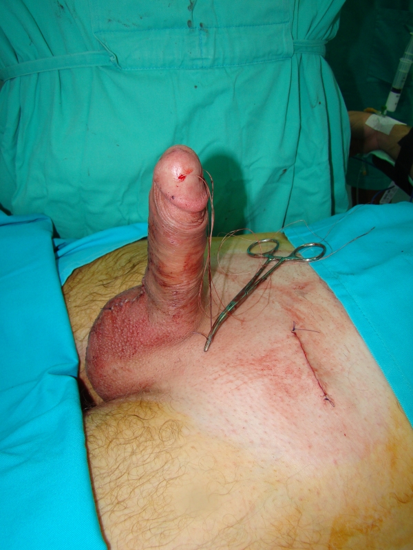
CASE 2
Peyronie’s disease – grafting technique

Severe dorsolateral curvature. Erection is preserved

All deformities are corrected using human pericardium graft
Appearance at the end of surgery. Erection is reestablished
CASE 3
Peyronie’s disease – grafting technique

Severe dorsolateral curvature. In erection, penis lies on anterior abdominal wall

Marked dorsal curvature is combined with hourglass phenomenon

Penile deformities are corrected

Wide neurovascular bundle completely covers tunical grafts

Appearance one month after surgery
CASE 4
Peyronie’s disease – grafting technique

Severe curvature verified by artificial erection

Tunica albuginea incision and opening

Penis is completely straightened and lengthened
CASE 5
Peyronie’s disease – penile prosthesis without grafting

Severe dorsolateral curvature

Semirigid penile prostheses are inserted using ventral approach

Tunica albuginea relaxing incisions are made to correct deformities

Wide neurovascular bundle completely covers tunical defects

Appearance at the end of the surgery

Outcome three months later
CASE 6
Maleable penile prosthesis for erectile dysfunction

Implantation of maleable prosthesis using ventral approach.

Result after penile prosthesis insertion.

Result after penile prosthesis insertion.
CASE 7
Inflatible penile prosthesis for erectile dysfunction

Insertion of penile prosthesis
Aspect at the end of the surgery

Outcome three weeks later

Outcome three weeks later
1. Kueronya V, Miernik A, Stupar S, Kojovic V, Hatzichristodoulou G, Egydio PH, Tosev G, Falcone M, De Luca F, Mulalic D, Djordjevic M, Schoenthaler M, Fahr C, Kuehhas FE. International multicentre psychometric evaluation of patient-reported outcome data for the treatment of Peyronie’s disease. BJU Int. 2015; 115(5):822-8.
2. Djordjevic ML, Kojovic V. Penile prosthesis implantation and tunica albuginea incision without grafting in the treatment of Peyronie’s disease with erectile dysfunction. Asian J Androl. 2013 May;15(3):391-4.
3. Djordjevic M, Majstorovic M, Bizic M, Kojovic V, Milosevic A, Pandey S. Penile prosthesis implantation and tunica albuginea incisions without grafting in the treatment of Peyronie’s disease. Europen Urology Video Journal, 2009; 15.3, (1).
4. Djordjevic M, Majstorovic M, Bizic M, Kojovic V, Milosevic A, Ivovic J, Zejnilovic N. Peyronie’s disease treatment by penile prosthesis implantation and tunica albuginea incisions without grafting. Urology, 2009;74(4, suppl 1) S159.
5. Perovic S, Djordjevic M, Sansalone S. Geometrical principle (egydio procedure) in the surgical treatment of Peyronie’s disease – our experience. Eur Urol Suppl, 2007;6(2): 286(ABS.1054).
6. Perovic S, Djordjevic M. Penile disassembly technique to surgical treatment of Peyronie’s disease. Br J Urol, 2001; 88: 731-739.
7. Basting R, Djakovic N, Djordjevic M, Perovic S. Surgical treatment of Peyronie’s plaque located under the glans cap using the penile disassembly technique and laser plaque treatment. Eur Urol, 2000; 37(suppl 2): 59.
8. Perovic S, Djordjevic M. Penile disassembly technique to surgical treatment of Peyronie’s disease. J Urol, 2000; 163(suppl.): 355.